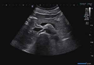
WHAT IS AN ULTRASOUND
 Ultrasound is a type of imaging that uses high-frequency sound waves to look at different organs inside the body. It is used to view the liver, kidneys, uterus, ovaries and numerous other organs and blood vessels. Ultrasound is also the desired modality to image a fetus during pregnancy.
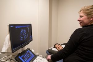
WHAT TO EXPECT WHEN YOU HAVE AN ULTRASOUND
During an ultrasound exam, you will be asked lie on an exam table, an ultrasound technologist applies a special gel and moves a device called a transducer over part of your body. The transducer emits sound waves, which will bounce back and produce an image on the monitor. The time it takes to complete each test will vary depending on which body part is being imaged. The typical exam time is between 30 and 60 minutes.
WHEN IS AN ULTRASOUND USED
Ultrasound can be used in different ways, depending on the type of test ordered and which part of the body is being examined.
Diagnostic medical sonography may be used to:
- Image arteries and veins in the body to find out if blood is flowing at a normal rate and level
- Check the thyroid gland for cancer or non- cancerous growths
- Check for abnormalities in the abdomen and kidneys
- Assist with needle guidance during a biopsy procedure, which removes a small sample of tissue for testing.
For women, diagnostic medical sonography may be used to:
- Image a lump or area of pain in the breast.
- Image the pelvic area to assist in finding the cause of pelvic pain, abnormal menstrual bleeding.
- Assist in diagnosing infertility or to monitor infertility treatments.
Pregnancy is a common indication for an ultrasound exam, and allows your doctor to get information about the health of an unborn baby.
- In the first trimester it is typically used to confirm pregnancy and estimate due dates and perform screenings for Down syndrome.
- Between 19-20 weeks your doctor may order an anatomy ultrasound, which will measure the fetus and check for any other abnormalities.
- In the third trimester an ultrasound may be used to track the growth, amniotic fluid, and position of the unborn baby.
Abdominal Ultrasound
 Examines internal organs, including the liver, gallbladder, spleen, pancreas, kidneys, and bladder. This can help to diagnose a variety of conditions and to assess the damage caused by illness.
For this exam you should have nothing to eat or drink for 8 hours prior to scheduled appointment.
Pelvic Ultrasound:
One of the more common forms of ultrasound, used to examine the uterus, ovaries, bladder, and prostate gland. Pelvic ultrasound is often used to diagnose conditions such as pelvic pain, abnormal bleeding, ovarian cysts, and uterine fibroids.
There are two types of Pelvic ultrasound:
- Transabdominal – An exam in which transducer is placed over the pelvic area to produce images.
- Transvaginal– A transducer with a protective cover and gel is inserted into the vagina and gently angled in order to obtain images.
For this exam you should drink 32 ounces of water at least one hour before your appointment. Do not empty your bladder until after the exam.
Generally, no fasting or sedation is required for a pelvic ultrasound, unless the ultrasound is part of another procedure.
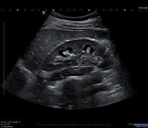
Obstetric Ultrasound
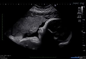 OB ultrasound refers to the specialized use of sound waves to visualize and determine the condition of a pregnant woman and her embryo or fetus. Obstetric ultrasound should be performed only when clinically indicated. Some indications may be:
- To establish the presence of a living embryo/fetus
- To estimate the age of the pregnancy
- To diagnose congenital abnormalities
- To evaluate the position of the fetus
- To evaluate the position of the placenta
- To determine if there are multiple pregnancies
Please note that we do not permit video recordings or photos from any recording devices (i.e. cell phone, camera, etc.) in the exam room. In addition, our ultrasound rooms are not large and we may need to limit the number of guests during your scan. There are many aspects of the pregnancy that the sonographer needs to assess during your ultrasound and therefore may limit discussion during the exam.
Nuchal Translucency:
A nuchal scan or nuchal translucency scan/procedure is a prenatal screening scan done during the first trimester. It’s a way of checking your baby’s risk of certain birth defects such as Down syndrome, Edward’s syndrome, and many other chromosomal abnormalities as well as heart problems.
The screening involves two steps. A blood test that checks for levels of two substances and an ultrasound that measures your baby’s nasal bone as well as the fluid at the back of your baby’s neck. A high volume of fluid can be a sign of problems.
A technician will take a quick blood sample from your arm or fingertip. The nuchal translucency screening is a normal ultrasound. You’ll lie on your back while a technician holds a probe against your belly. It will take between 20 to 40 minutes.
Renal Ultrasound
 Renal ultrasound determines the size, shape, and function of the kidneys, and can be useful in the detection of kidney stones, cysts, and tumors.
For this exam you should drink 24 ounces of water prior to exam and hold.
Vascular Ultrasound
Vascular ultrasound is a fast, noninvasive way of identifying issues and analyzing the flow of blood through the arteries and veins. These images can help the physician to see and evaluate vascular problems such as blockages, clots, plaque and congenital malformations
Thyroid Ultrasound:
Thyroid Ultrasound checks for underactive and overactive thyroid glands, nodules, and cysts.
Ultrasound Pyloric Stenosis
An ultrasound done to rule out or diagnose Pyloric Stenosis in infants. Pyloric Stenosis is a condition in which the opening between the stomach and small intestine thickens.
Pyloric stenosis occurs most often in babies under six months. In this condition, the pylorus muscles block food from entering the small intestine. It occurs more often in males.
Pyloric stenosis can lead to forceful vomiting, dehydration, and weight loss. Babies with this condition may seem to always be hungry.
For this exam there is nothing to eat or drink for 4 hours. Bring a bottle of Pedialyte to drink during the exam.
Ultrasound Mesenteric Artery
A mesenteric artery ultrasound is a non-invasive imaging procedure that uses high-frequency sound waves to produce images of your mesenteric arteries, the arteries that supply oxygenated blood to your digestive organs. This test helps to detect blockages or narrowing of the arteries in the abdominal area.
For this exam please bring 2 bottles of Ensure to drink during your study.
Ultrasound guided biopsies:
- Breast